Thermography – Medical Infrared Thermal Imaging (MTI)
With each breath we take, most of us are not aware of the constant, pulsating cellular activity that literally keeps us alive and is constantly challenged to keep us healthy. It is easy for us to ignore the invisible physiological signs of cellular and metabolic stress. The astounding truth – this is the level where all abnormalities begin and also the level where abnormalities are easiest to correct. As cellular and metabolic stresses cascade into progressive degeneration, they ultimately reveal themselves in the manifestation of anatomical damage many years later. Today, patients and doctors find themselves with late stage detection requiring aggressive treatment.
The phenomenal truth is that we can detect the changes at these early stages through the lens of the Medical Infrared Thermal Imaging (MTI) camera. Thermography provides a crucial entryway to preventive medicine as a method of controlling the diseases that we currently only recognize at the latest advanced stages.
What You Should Know About Thermography
Medical Thermography is the use of an infrared camera to “see” and “measure” what we call metabolism, thermoregulation or thermal energy emitted from the body. Thermography reveals a fascinating and reliable pattern of thermal activity that discloses a silent warning. These patterns can see the root cause of such conditions as: headaches, allergies, dental pathologies, carotid artery disease, breast cancer, digestive dysfunction, liver/gallbladder disease, musculoskeletal conditions, pain, and immune dysfunction. It is most well researched for its ability to detect risks for breast cancer much earlier than any other screening tool. In fact, studies have shown that it can detect early signs of breast cancer up to 8 years before a mammogram. A sensitive infrared video camera can even detect a gentle but visible pulsation created by blood pumping through blood vessels.
Infrared lets us see what our eyes cannot because it’s part of an invisible electromagnetic wavelength that we perceive as heat. Everything with a temperature above absolute zero emits heat. Infrared thermography cameras provide us with the ability to record precise non-contact body temperature measurement. Abnormal physiological activities in the body are easily observed with thermography. This makes infrared cameras an extremely cost-effective, safe and valuable diagnostic tool for determining a patient’s general health picture for many applications including breast and cardiovascular monitoring.
Thermography technology has been prevalent for more than 50 years (the first thermal image was taken in 1952); the FDA approved and registered the first medical thermography system over 20 years ago. The medical application enjoyed a period of popularity in the 70’s; it has now emerged again in recent years since advancements in the technology has made the imaging more reliable resulting in favorable population acceptance. Even though it is classified by many insurance companies as “investigational” or “experimental,” the practical reality is that the medical evidence consistently supports that MTI has advanced far beyond either of these imposed labels. More and more of the public is asking for this type of screening tool especially for breast cancer prevention.
When Should You Use Thermography
Thermography is most useful in the following areas:
Inflammatory Phenomena- includes early detection of cardiovascular disease, arthritis, fybromyalgia or trauma such as strains, sprains, chronic pain or infection.
Neovascular Phenomena – cancer is fed by the bodies own blood supply; this development of early vascularity can be detected well before anatomical changes which are detected with other screening tools; MTI can detect these early changes and cue us into taking preventive action.
Natural approaches to improve the evolving condition.
Neurological Phenomena – chronic regional pain syndrome and nerve irritation can cause referred pain in other areas. Circulatory deficits are easily seen in thermographic images.
How Does Thermography Compare And Compliment Mammography
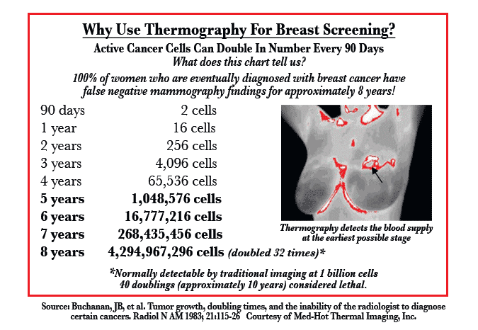
The U.S. has the highest rate of breast disease in the world. The good news is that research has finally confirmed that it is not primarily related to family history (less than 5% of all breast cancers are genetically related). Therefore, it is a PREVENTABLE disease. That means that you can develop a strategy for minimizing your odds of developing breast disease by making basic lifestyle changes while incorporating new, predictive screening technologies like MTI. FDA approved, Thermography can forewarn women of breast health problems up to 8 years before symptoms may be visible. It is much better to detect breast cancer than simply avoid it. The following chart clearly demonstrates the extreme value of thermography:
This is a hypothetical chart, but it represents the average growth pattern of a slow-developing breast tumor. Most doctors agree and even tell their breast cancer patients that a growth may have been there for 8 or 10 years.
Mammograms are a good tool for determining the exact location of a developed tumor, but it is not an early warning system. “Early” is a relative term, so if a mammogram can see a tumor in the 8th year, it is earlier than the 10th year, but in any case, even the 7th year may be too late to change the outcome.
The real danger of breast cancer is determined by whether or not the tumor has spread to a vital organ. The longer the tumor exists, the longer it has to spread. We now have earlier detection through MTI!
Thermography can see the blood supply that feeds a tumor in its microscopic infancy, and the only way to possibly detect it in that stage is to establish a thermographic baseline and monitor it every year for the real early signs. MTI has been comprehensively researched, and while it cannot be called a replacement for Mammography, it has many valuable advantages including: earlier detection of suspicious patterns; an adjunct to inconclusive mammograms; improved detection for women with dense breasts or implants; and a reasonable alternative for women who refuse mammogram. Below is a sample of the over 800 studies in the index-medicus; they represent some of the important findings and value of thermography.
In 1982, the FDA approved breast thermography as an adjunct diagnostic breast cancer screening procedure. Of the extensive research conducted since the late 1950’s, well over 300,000 women have been included as study participants; the size of the studies are very large: 10k, 37k, 60k, 85k. Some studies have followed participants up to 12 years.
Strict standardized interpretation protocols have been established for 15 years to remedy problems with early research.
Breast thermography has an average sensitivity and specificity of 90%.
An abnormal thermogram is 10 times more significant as a future risk indicator for breast cancer than a first order family history.
A persistent abnormal thermogram carries with it a 22x higher risk of future breast cancer.
Extensive clinical trials have shown that breast thermography significantly augments the long term survival rates of its recipients by as much as 61%.
When used as a multi-modal approach (clinical exam + mammography + thermography), 95% of early stage cancers will be detected.
Our patient care process combined with our state-of-the-art technology results in a system that far surpasses any other screening center in Connecticut.
Our Thermography screening is unique for these reasons:
Images are analyzed by specially-trained MDs – some of the most qualified interpreters in the industry.
We not only screen clients, but can also provide natural treatment solutions for conditions uncovered using Thermography with various natural medicine tools and techniques.
Thermography is described in part by the FDA as “adjunctive diagnostic screening for detection of breast cancer or other uses”. Thermography is not a stand-alone device and does not replace any other diagnostic device or examination.
Kenneth R. Hoffman D.Ac.(RI), L.Ac., CCH
Medical Director
This information, including but not limited to opinions and/or recommendations contained herein are for general educational informational purposes only. Such information is not intended to be a substitute for professional medical advice, diagnosis or treatment. Copyright 2015, SNHC

